
Major points
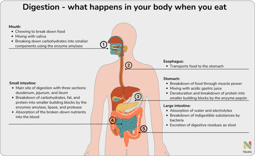
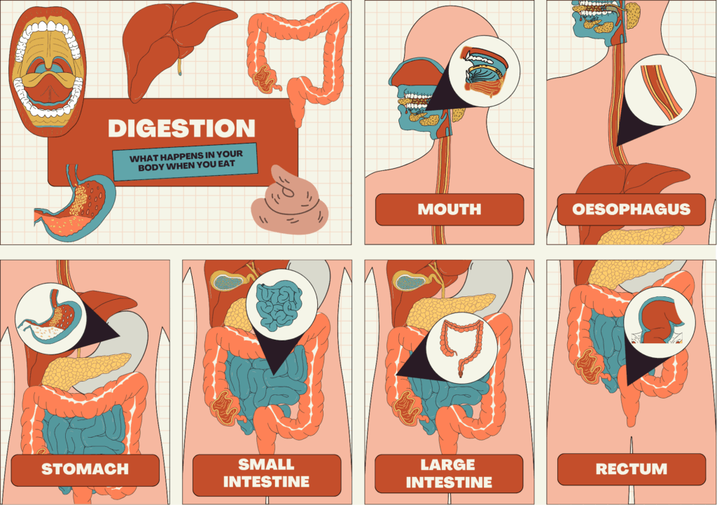
- Digestion begins in the mouth. Food is broken down, mixed with saliva, and transported through the esophagus to the stomach.
- Most of the digestive process happens in the stomach and small intestine. Nutrients are broken down by enzymes and absorbed into the bloodstream.
- The large intestine removes water from the remaining food matter, bacteria break down leftovers, and waste is excreted.
Mouth – start of digestion
We all eat multiple times a day – usually in the morning, at noon, and in the evening, plus one or two snacks. Eating is essential. Frequency is more of a personal preference. Some people fast for several hours a day and eat only at specific times, while others eat whenever their body signals that it is hungry. But have you ever asked yourself what exactly happens in your body after you eat?
The process of taking in solid or liquid food through the mouth and breaking it down into small molecules so the body can absorb nutrients is called digestion.
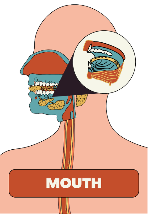
Food is taken in through the mouth. The teeth mechanically break down the food into smaller pieces. Smelling the food, chewing, and tasting stimulate the salivary glands to produce saliva. About 1.5 liters of saliva are produced each day. This saliva contains the digestive enzyme α-amylase, which starts breaking down starch – such as in potatoes or bread – into disaccharides right in the mouth. The more thoroughly food is chewed, the greater its surface area becomes, allowing digestive enzymes to work more effectively. Saliva also changes the consistency of food into a soft mass, making it easier to glide through the esophagus. The tongue shapes the chewed food into a bolus and pushes it into the throat. This triggers the swallowing reflex. The better the food is broken down through chewing, the easier it can pass through the entire gastrointestinal tract.
The taste perceived while chewing also signals the stomach glands to release gastric juice, preparing the stomach for the arrival of food.
Esophagus – transporting the food
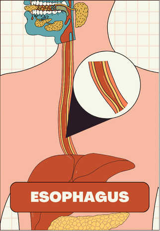
The esophagus is a muscular tube about 25 centimeters long that transports food from the mouth to the stomach. The food bolus is moved to the stomach through wave-like muscle movements called peristalsis. To prevent food from entering the windpipe, the epiglottis closes off the trachea. The lining of the esophagus produces mucus to help food slide more easily. At the lower end of the esophagus is a sphincter muscle that opens to allow food into the stomach and then closes again to prevent stomach acid from flowing back. If acid does make its way back into the esophagus, it can cause burns that are painful and may permanently damage the esophageal lining. Frequent acid reflux can lead to inflammation of the esophagus, known as reflux esophagitis, which over time increases the risk of complications such as scarring, narrowing of the esophagus, or even esophageal cancer.
Stomach – mechanical and chemical processing of food
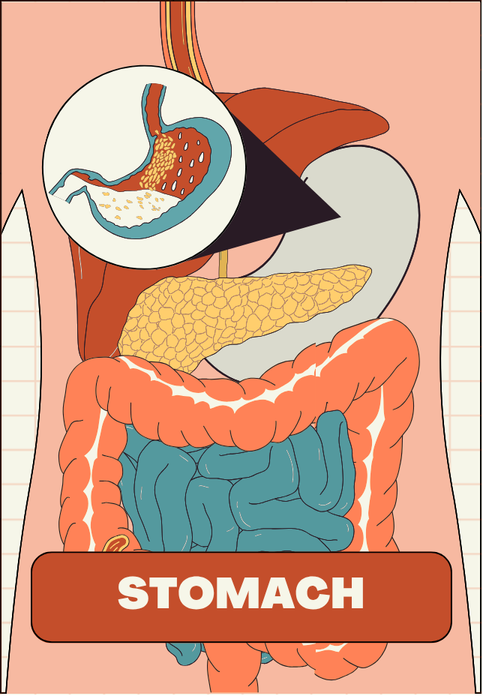
The stomach first acts as a temporary storage container and is lined with a mucous membrane. The food is kneaded by wave-like movements of the stomach muscles and mixed with gastric juice. About 2 liters of gastric juice are produced throughout the day by specialized cells in the stomach lining. Gastric juice contains hydrochloric acid, mucus, and pepsinogen, which is the inactive precursor of the enzyme pepsin. Hydrochloric acid plays several important roles: it kills pathogens, denatures proteins (changes their structure to make them easier to break down), and activates pepsinogen into pepsin. Pepsin then breaks down large protein molecules into smaller ones. The stomach’s mucous cells produce an alkaline mucus containing mucin. This protects the stomach lining from the aggressive hydrochloric acid.
The entire process gradually transforms the food into a thick, semi-liquid mass known as chyme. The stomach releases the chyme in small portions through the pyloric sphincter into the small intestine. Depending on its composition, food can remain in the stomach for two to six hours. A high fat content slows stomach emptying, while well-chewed, sugar-rich, and liquid foods pass through more quickly.
Small intestine – main site of digestion and nutrient absorption
The small intestine is about 5 to 6 meters long. Its role is to mix the chyme, continue digestion, and move it toward the large intestine through peristaltic movements. The lining of the small intestine produces about 1 to 2 liters of digestive juice daily. This digestive juice also contains the mucous substance mucin, which protects the intestine from stomach acid and other harmful substances. The small intestine is coiled and made up of three sections:
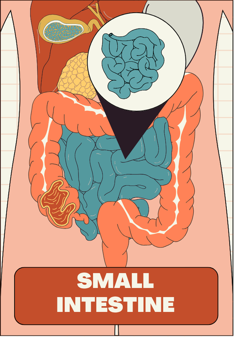
- The duodenum is the first section. This is where the acidic chyme from the stomach is neutralized. This neutralization is carried out by alkaline juice produced by the pancreas. About 1.2 to 1.5 liters of pancreatic juice are produced daily. At the same time, essential digestive enzymes such as amylases, lipases, and proteases are released into the duodenum by the pancreas. These enzymes break down carbohydrates, fats, and proteins into smaller units. In addition, bile acids produced by the liver and stored in the gallbladder are released into the duodenum. These emulsify fats, meaning they break down large fat molecules into smaller droplets. This gives the enzyme lipase a larger surface area to split triglycerides into glycerol and free fatty acids, which are then absorbed through the intestinal wall. Without emulsification, only a small portion of fat molecules could be absorbed.
- The jejunum is the second section. Here, the chyme from the duodenum is further broken down by enzymes, and most nutrients are absorbed into the bloodstream through the intestinal wall. To ensure maximum nutrient absorption, the lining of the jejunum has villi (finger-like projections) and microvilli (microscopic extensions), which increase the surface area to as much as 200 square meters. This is where the broken-down macronutrients – carbohydrates as simple sugars, proteins as amino acids, and fats as glycerol and free fatty acids – are absorbed. Fat-soluble vitamins A, D, E, and K, as well as water-soluble vitamin C, all B vitamins (except B12), and minerals such as calcium, chloride, potassium, magnesium, and sodium are also absorbed here.
- The ileum is the final section. Its main job is to absorb nutrients not yet taken up previously. It is the only part of the small intestine that absorbs vitamin B12 and reabsorbs bile acids so they can be reused. The ileum also has villi and microvilli and absorbs water and electrolytes, further thickening the remaining chyme. Up to 8 liters of fluid per day -coming from drinks, food, and digestive secretions (saliva, gastric juice, pancreatic juice, bile, intestinal juice) – are reabsorbed here. All components that are not absorbed by this point, such as indigestible fiber, are passed on to the large intestine.
Large intestine – water absorption and processing leftovers
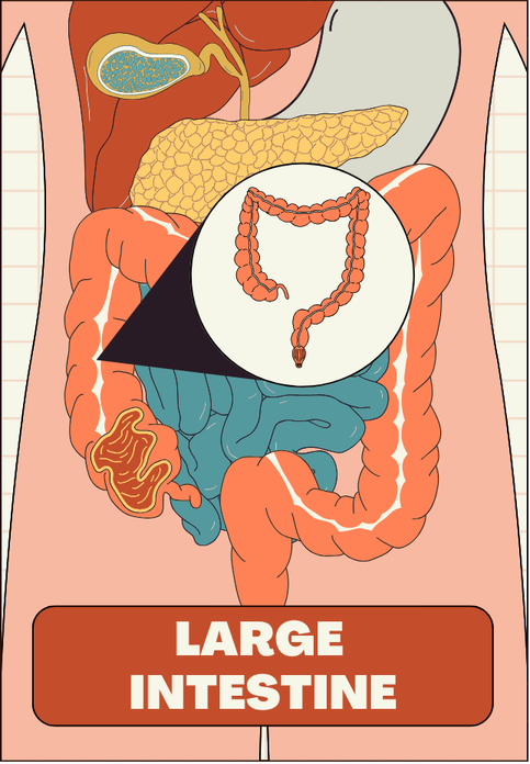
The large intestine primarily absorbs water and electrolytes like potassium and sodium from the remaining chyme. When chyme arrives from the ileum, it still contains about 0.5 to 1.5 liters of water and indigestible parts such as fiber. The large intestine removes most of the water, thickening the mass into stool. Unlike the small intestine, the large intestine has no villi but has crypts – indentations that produce mucus. Stool is mixed with this mucus to make it easier to pass. Compared to the rest of the gastrointestinal tract, peristalsis here is slower. The intestinal content is moved only every four to six hours. In the meantime, the content is moved back and forth by antiperistalsis – reverse peristaltic movements – allowing for more water to be absorbed.
A healthy large intestine hosts around 400 different bacterial species, known as the gut microbiome. These bacteria continue breaking down water-soluble fiber, producing gases, and short-chain fatty acids. The large intestine is, therefore, not only responsible for water absorption but also plays a role in processing food components that were not broken down in the small intestine. Nutrients produced by these bacteria enter the bloodstream, while the indigestible remains, bacteria, and dead cells are passed into the rectum as stool.
Rectum and anus – excretion
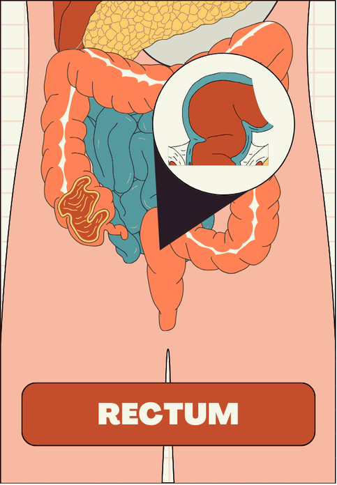
The digestion process ends in the rectum, where stool is temporarily stored before excretion. Each time stool arrives, the rectum stretches. Specialized nerves monitor pressure and fullness. Once the rectum is filled, a reflex is triggered that signals the body to prepare for a bowel movement.
Excretion occurs when the person is in the proper position and consciously opens the external sphincter. About 100 to 200 grams of stool are excreted daily through the anus.
During digestion, the broken-down macronutrients are reassembled in your cells and can then be used as energy stores. This process is called anabolism. You can learn more about anabolism, catabolism, and metabolism in general here.
Bibliography
Bundesministerium für Ernährung und Landwirtschaft: Gutes Bauchgefühl. Gesundheit beginnt im Darm. Kompass Ernährung (3/2021), checked on 2/6/2025.
Bundeszentrum für Ernährung (Ed.) (2025): Der Weg der Nahrung. With assistance of BZfE Larissa Kessner, Bielefeld Silke Hoffmann. Bundeszentrum für Ernährung. Available online at https://www.bzfe.de/schule-und-kita/material-fuer-die-schule/sekundarstufe/der-weg-der-nahrung, updated on 1/22/2025, checked on 4/9/2025.
Dr. Lioba Hofmann (2017): Nahrung und Verdauung. Wissen kompakt. With assistance of Larissa Kessner BLE. Auflage 3.